A fibroadenoma is the most common benign lump in the female breast. It is made up of connective and mammary gland tissue that feels like rubber. However, in very rare cases, a fibroadenoma becomes cancerous. Find out more about the treatment options, symptoms, diagnosis and causes of fibroadenoma here.
ICD codes for fibroadenoma: D24
Quick overview
• Symptoms: A fibroadenoma usually causes no symptoms. Sometimes it leads to breast tenderness or a changed breast shape, among other things.
• Causes and risk factors: Female sex hormones (estrogens) stimulate the proliferation of fibroadenoma cells.
• Diagnosis: Various examination methods are available for diagnosis, including sonography, mammography, magnetic resonance imaging and tissue examinations.
• Treatment: The treatment of a fibroadenoma is not mandatory. In some cases, however, surgical removal makes sense.
• Disease course and prognosis: Fibroadenomas that are not removed may remain the same size. They very rarely develop breast cancer.
• Prevention: There are no clear recommendations to prevent the development of a fibroadenoma.
What is a fibroadenoma?
Benign connective tissue knots are generally referred to as fibroids. These are benign tumors that occur in breast tissue, among other things. If such a fibroma in the breast also contains glandular tissue, it is referred to as a fibroadenoma.
Fibroadenomas are the most common benign breast (mammary) lumps in women between the ages of 20 and 40 and are most commonly found in women of childbearing age. However, they also occur in older women going through the menopause and in women undergoing hormone treatment. If women take the pill, however, they develop less frequently.
A fibroadenoma arises from mammary gland and connective tissue and feels tough like rubber. Fibroadenoma pain sufferers usually do not feel. You often discover the knot by accident when it is an inch or two in diameter.
It is usually a single nodule, more rarely multiple fibroadenomas. In five to ten percent of cases, both breasts are affected.
Only rarely does the fibroadenoma become larger than three centimeters. It may even go away on its own with age. In young women, however, there is a special form that grows very quickly and may change the shape of the breast.
Since it is a benign lump, removal of the fibroadenoma is not mandatory from a medical point of view. In some cases, however, and at the patient’s request, the knot is surgically removed.
Can a fibroadenoma become malignant?
Very rarely (in less than one percent of those affected), a mammary carcinoma (breast cancer) develops from the fibroadenoma. Affected women are nevertheless recommended to have their breasts checked regularly by a gynaecologist.
Disease course and prognosis
A fibroadenoma is a benign tumor. If not removed, it may remain the same size.
During menopause, the proportion of female sex hormones in the body decreases, so that a fibroadenoma may regress by itself. On the other hand, if the hormone level increases, for example during pregnancy or under hormone therapy, the fibroadenoma may become larger. Nodes that are incompletely removed may grow back (recurrence).
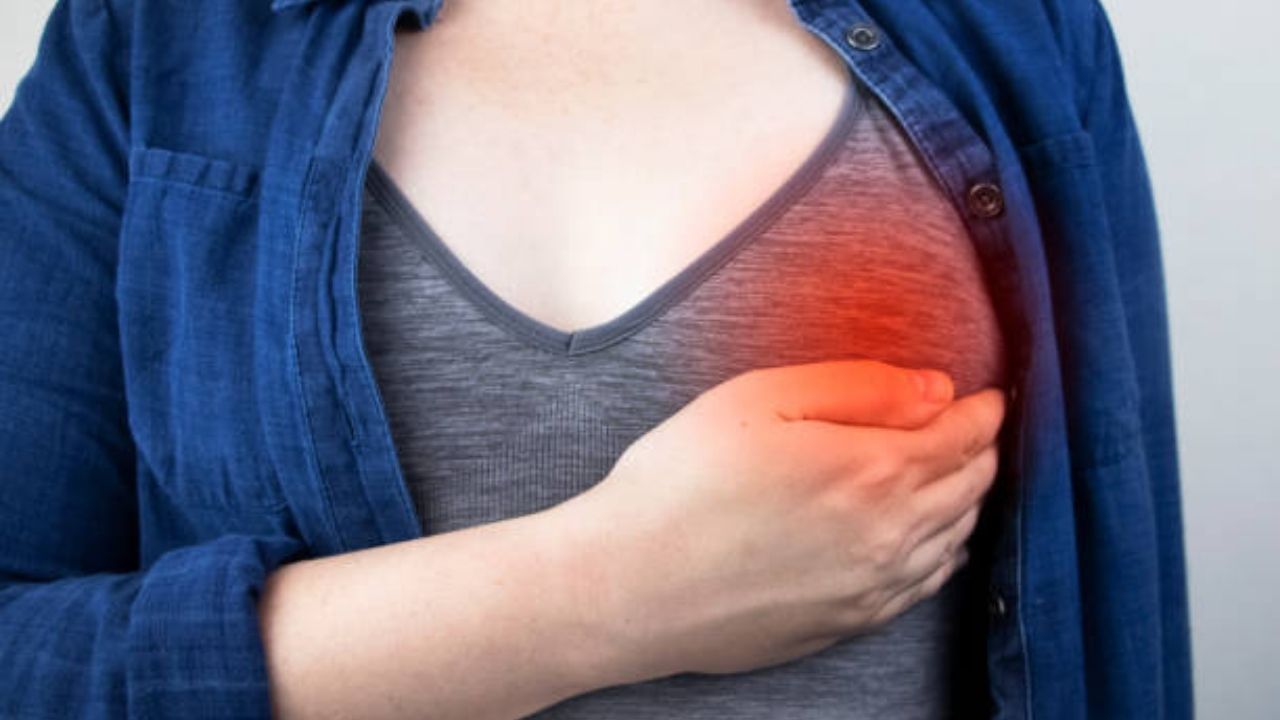
Symptoms of fibroadenoma

The fibroadenoma is mostly asymptomatic. Women discover a rough lump in the breast more by accident. Sometimes there are several knots close together. When the gynecologist touches the breast (mammary) during the annual check-up, almost no woman feels pain from the fibroadenoma.
Sometimes fibroadenoma breasts can feel tight, especially before your period. Some women find this uncomfortable or slightly painful.
In rare cases, and mostly in younger women, there is a fast-growing type of this lump. Then the fibroadenoma may change the shape and size of the breast. The breast may bulge slightly as a result and appear enlarged.
Causes and risk factors of fibroadenoma
Female sex hormones (estrogens) stimulate fibroadenoma cells to multiply. If the concentration of these hormones increases , for example under hormone treatment or during pregnancy, many new glandular and connective tissue cells are formed. In some cases, these then form a tough knot, the fibroadenoma.
Diagnosis of fibroadenoma
Most women notice fibroadenoma themselves when they examine their breasts. In order to make the diagnosis, the doctor first asks about symptoms and the previous history (anamnesis).
It is important that the doctor excludes that it is a malignant change (breast cancer) in contrast to a fibroadenoma. He checks whether the shape of the breast has changed and feels the breast and armpits. If he discovers a conspicuous knot or another unclear change, the following examinations may follow:
Sonography (ultrasound)
A fibroadenoma can usually be adequately identified by ultrasound and can be differentiated from other nodes. The doctor applies gel to the breast and armpit and systematically scans the breast with the ultrasound probe. This exam is not painful. At most, the cold ultrasound gel may be perceived as uncomfortable.
Mammography
This is a special X-ray examination that is carried out from the age of 50 for regular early detection of breast cancer.
For the X-ray, the breast is pressed between two plates. Most women find this harmless procedure painful. However, it is necessary to spread apart the healthy mammary gland tissue and distinguish a malignant lump from a fibroadenoma.
In most cases, this examination results in a further diagnosis. Mammography is not suitable for young women. Your breast tissue is still so dense that changes are difficult to identify. For women over the age of 40, however, mammography is the preferred examination method.
Magnetic resonance imaging (MRI)
In certain cases, magnetic resonance imaging or magnetic resonance imaging is necessary to diagnose or rule out a fibroadenoma. This includes women who have had breast surgery, have silicone implants, or have had breast cancer in the past.
Changes in dense glandular tissue can also be clearly seen on MRI. For the examination, patients sometimes have a contrast agent injected into a vein. During the recording in the examination tube, you must lie as still as possible. Magnetic resonance imaging does not produce any harmful radiation.
Punch biopsy
Despite the examination options mentioned, it is sometimes not possible to obtain a clear result. If necessary, the doctor then resorts to a punch biopsy for diagnosis.
This examination is also carried out if one of the other examinations has led to a conspicuous result and needs to be clarified further. Using a type of gun (similar to earring shooting), the doctor removes a cylindrical piece of tissue from the knot.
The breast is usually anesthetized locally beforehand so that there is as little pain as possible. The tissue sample is then examined under a microscope by a tissue specialist (pathologist). This is how he determines whether it is a fibroadenoma or another node.
How is a fibroadenoma treated?
If a fibroadenoma has been diagnosed, treatment is not absolutely necessary from a medical point of view. It is only recommended to have the breast examined regularly by the gynaecologist. In this way, he quickly recognizes when the fibroadenoma has turned malignant and needs to be treated in another way.
However, if the lump grows very quickly and changes the shape of the breast, many women choose to have the fibroadenoma removed. If someone in the family has breast cancer, this minor operation is also recommended.
Depending on where the fibroadenoma is and how big it is, the surgery may change the shape of the breast. Also, some fibroadenoma cells sometimes remain in the breast despite the operation. These cells may then form a node again.
Due to the hormonal changes during pregnancy, fibroadenomas may grow faster and cause symptoms. For this reason, doctors often recommend removing fibroadenomas before pregnancy. After the procedure, most mothers are able to breastfeed without any problems. However, breast engorgement occasionally occurs.
Node removal (excision)
For larger fibroadenomas, rapid growth, and women older than 40, the doctor usually surgically removes the entire lump and has it examined in a laboratory.
Prevention of fibroadenoma
There are no clear recommendations on how to prevent the development of fibroadenomas (e.g. through a targeted diet). However, women have the opportunity to contribute to the early detection of fibroadenoma by regularly examining their breasts themselves and attending their check-up appointments.
Even after the surgical removal of a fibroadenoma, those affected are recommended to examine their breasts thoroughly once a month, shortly after menstruation. In this way, nodules that develop from any remaining fibroadenoma cells can be detected at an early stage.