Fibrous dysplasia is a mostly congenital malformation of the bones. It occurs mainly in children and adolescents. Instead of growing properly, individual bones only form fibrous growths. Read everything you need to know about this disease here.
ICD codes for this disease: Q78 | M85
Quick overview
• Prognosis: Generally good, course often ends at the end of puberty; the severe and very rare form of McCune-Albright syndrome can also be treated.
• Causes and risk factors: Non-hereditary mutation of a specific gene (GNAS gene) on chromosome 20, cause has not yet been researched, usually occurs before birth, sometimes only after birth.
• Symptoms: If only one bone is affected, the disease often remains without symptoms, otherwise bone pain among other things; various symptoms in the very rare McCune-Albright syndrome, in addition to bone deformities, including premature puberty and pigment disorders.
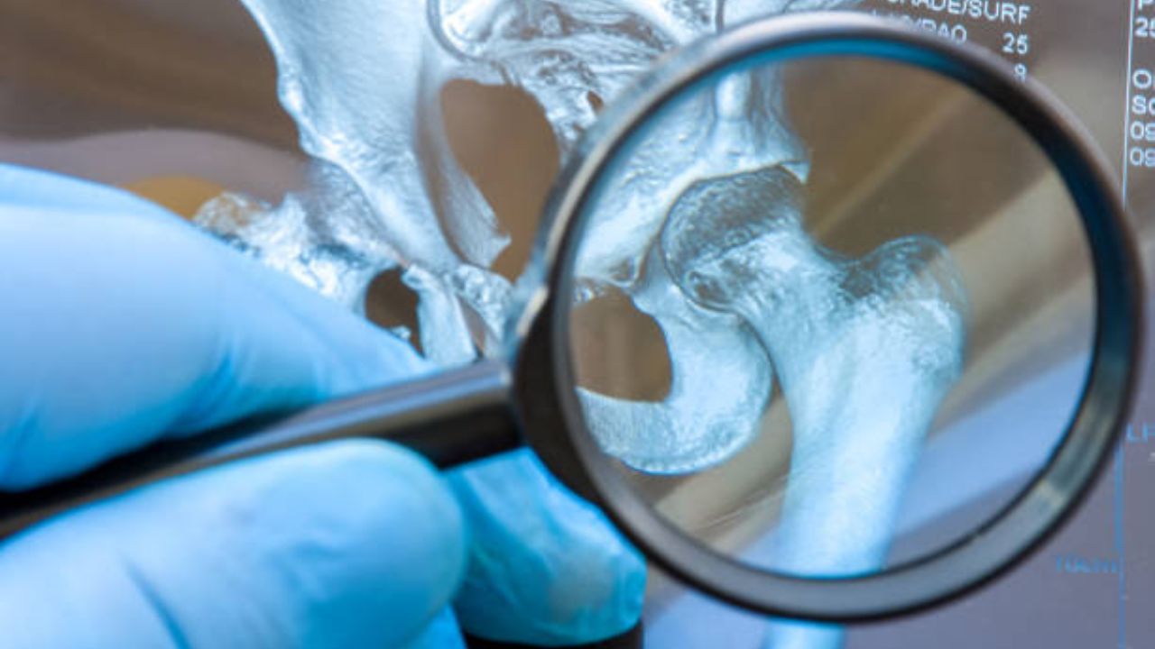
• Diagnostics: X-ray, computer tomography, tissue samples and other examinations if several bones are affected.
• Treatment: Depending on the severity and location, treatment of symptoms; splints of affected bones, physiotherapy, surgical removal of bone growths; in McCune-Albright syndrome treatment of other symptoms; Treatment of the causes is not yet possible.
What is fibrous dysplasia?
Fibrous dysplasia is a condition, usually congenital, in which one or more bones fail to develop in their normal cellular structure; instead, tumors arise from fibrous connective tissue. This most commonly occurs in a femur, but the tibia, ribs, skull and jaw bones are also affected relatively often.
Fibrous dysplasia is based on a genetic defect, but is not hereditary. Males and females are equally affected, with progressive forms with multiple affected bones (polyostotic fibrous dysplasia or Jaffé-Lichtenstein syndrome) appearing to be more common in girls. Children and adolescents between the ages of five and 15 are most frequently affected, and adults are more rarely affected.
Although one of the most common bone diseases, fibrous dysplasia is a rare disease overall. Exact figures are not known, since in many cases fibrous dysplasia progresses without noticeable symptoms; Doctors therefore assume that it often goes undiagnosed.
Fibrous dysplasia: Different manifestations
Doctors distinguish different forms of the disease:
• Monostotic fibrous dysplasia (70 percent): only one bone is affected.
• Polyostotic fibrous dysplasia (25 percent): multiple bones are affected (Jaffé-Lichtenstein syndrome).
• McCune-Albright syndrome (very rare): fibrous dysplasia with “café au lait spots” (pigmentation disorder) and premature sexual maturity.
McCune-Albright syndrome is often listed as a separate condition, but shares the same genetic basis as fibrous dysplasia.
Is fibrous dysplasia curable?
Fibrous dysplasia has a good prognosis. The course varies from case to case. In some patients, the foci increase in stages during puberty, so that the affected bones are further distended. As a rule, however, no new herds develop. By adulthood at the latest, the fibrous dysplasia usually comes to a standstill and the bone is no longer remodeled. Three out of four affected patients are under 30 years old.
It is very rare for fibrous dysplasia to develop into a malignant bone tumor (osteosarcoma), occurring in less than one percent of cases. Severe courses are just as rare, for example when the fibrous dysplasia affects the skull bones and these are deformed in such a way that they limit the function.
The very rare progressive form of McCune-Albright syndrome with many affected bones, premature puberty and a large number of other possible symptoms can basically be treated according to the symptoms. Without therapy, those affected may have a reduced life expectancy.
If fibrous dysplasia is treated early, those affected have no restrictions on their quality of life.
Causes and risk factors
Fibrous dysplasia is due to a change in the genetic material: the so-called GNAS gene (guanine nucleotide-binding protein) of the 20th chromosome is changed (mutated). This causes the enzyme adenylyl cyclase to produce too much cAMP (cyclic adenosine monophosphate), a specific substance that plays an important role in signaling in the cell.
Ultimately, the mutation means that the spongy inner layer of the bone – the so-called spongiosa – is not properly formed. In their place is a soft, non-mineralized, connective tissue-like bone substance (osteoid). The cells divide before they are properly differentiated, which often leads to bone swelling.
The genetic defect only affects body cells and not the germ cells (sperm and egg cells) – this is referred to as a somatic mutation. The gene probably mutates during embryonic development or in early childhood. The mutation is therefore not in the genome from the start. This means that the GNAS gene defect is not hereditary. Why this mutation occurs has not yet been conclusively researched. The mutation sometimes occurs before birth, sometimes only after birth.
How is fibrous dysplasia manifested?
Fibrous dysplasia progresses very differently. Therefore, symptoms vary depending on the severity and which bones are affected. While some of those affected are completely symptom-free, others show various symptoms:
• Slight pulling bone pain.
• Stress pain (e.g. in the case of an affected femur).
• Difficulty walking, causing some people to limp.
• Frequent spontaneous bone fractures with actually light loads (fatigue fractures).
• Externally visible “bumps”, curvatures, and other changes in the bones (such as a visibly deformed facial skull).
• Rapid physical development in affected children and adolescents (rapid growth and early puberty).
• Pigment disorders, so-called café-au-lait spots.
The last two symptoms belong to the so-called McCune-Albright syndrome ; very few people with fibrous dysplasia are affected. “Café-au-Lait spots” are uniform, light brown, sharply defined spots that also occur, for example, in the hereditary disease neurofibromatosis type 1 (Recklinghausen’s disease). In McCune-Albright syndrome, they are often concentrated on one side of the body – just like the fibrous dysplasia or the areas affected.
The early onset of puberty is due to a change in the hormone balance. Sometimes fibrous dysplasia occurs together with other hormonal diseases, such as diabetes, Cushing’s disease or an overactive thyroid.
Depending on the region affected, the fibrous growths press on nerves or blood vessels in or on the bones, causing symptoms such as pain or circulatory disorders.
In addition to the bone deformations in fibrous dysplasia, there are rarely functional disorders of organs such as the heart, liver, pancreas or other organs if benign tumors, so-called myxomas, form in these.
Commonly affected bones
Basically, fibrous dysplasias are possible in all bones, but they occur particularly often in the following regions:
- Skull bones.
- Face, often the jaw.
- Ribs.
- Upper arm.
- Hips.
- Thigh.
- Shin.
Fibrous dysplasia: investigations and diagnosis
To diagnose fibrous dysplasia, the doctor will first ask about the symptoms and medical history. For example, he asks about possible pain, in which situations it occurs and whether one or more areas are affected. During a physical exam, he looks at the abnormal spots and feels them to check for changes.
The doctor may take blood from the affected person. In the case of fibrous dysplasia, normal levels of calcium and phosphate appear in the blood serum; instead, the value for the enzyme alkaline phosphatase is often increased. This blood value belongs to a group of enzymes that, among other things, often indicate changes in bone metabolism.
The main method to diagnose fibrous dysplasia is x-ray examination. It is not uncommon for an X-ray to raise suspicion of the rare disease, for example after a broken bone. The doctor often sees a spindle-shaped, opaque, thickened area in the affected bone, where the bone structure has been replaced by connective tissue. Some of the changes look like bone cysts.
The outer layer of bone (cortex) is usually thinner than in healthy bone. Computed tomography (CT) helps the doctor to take a closer look at the changes. The person being examined lies in a special device that takes very precise X-ray images of the body layer by layer in the form of sectional images.
Another imaging technique is skeletal scintigraphy. With this nuclear medicine examination, changes in the bone metabolism can be displayed. If polyostotic fibrous dysplasia is suspected, skeletal scintigraphy helps to identify all affected bones (foci).
Especially when only a single bone is changed (monostotic fibrous dysplasia), the correct diagnosis using imaging methods is sometimes difficult, since some other diseases present themselves in a similar way (e.g. bone cysts, benign fibrous histiocytoma, hemangioma, chondrosarcoma). In this case, the doctor takes a tissue sample from the changed area (biopsy), which is then examined under the microscope.
In the case of fibrous dysplasia, a typical structure of connective tissue containing collagen of different densities appears, interspersed with small trabeculae of bone tissue (so-called bone trabeculae). There are only a few “primordial bone cells” (osteoblasts) on their surface, but collagen fibers arranged in a radial pattern.
Treatment of fibrous dysplasia
A causal treatment is not possible with fibrous dysplasia. If the femur or shinbone is affected, it makes sense to relieve the bone, for example with a splint, depending on the case. This prevents possible fractures in the unstable areas.
It is best for those affected to discuss this with an experienced orthopaedist, as bone deformation sometimes occurs during growth. Physical therapy with strength exercises will help strengthen the surrounding bones and muscles.
If the fibrous dysplasia causes severe pain, this is usually contained with painkillers (analgesics). A relatively new therapeutic approach are so-called bisphosphonates – drugs that are also used to treat osteoporosis and other bone diseases. They appear to have a beneficial effect on the bone pain and fractures that accompany fibrous dysplasia and slow the progression of the disease.
In rare cases, fibrous dysplasia is so pronounced that, for example, bones in the face are severely deformed. Then an operation may make sense for aesthetic reasons.
The surgeon now plans the operations using modern 3D imaging and modeling methods, so that sensitive structures such as nerves and blood vessels are protected during the procedure.
Prevention
Fundamental prevention of fibrous dysplasia is not possible, since the actual cause of the gene mutation is not yet known. However, specific symptoms such as broken bones caused by splints on the weakened bone structure or McCune-Albright syndrome can be prevented by drugs that inhibit premature sexual maturity.