The term skin cancer includes various malignant skin diseases. The most common is “white skin cancer”, followed by the much more dangerous black skin cancer. Skin cancer symptoms and treatment depend on the type of cancer. Here you can read more about the topic: What does skin cancer look like? What are the causes? How is it diagnosed and treated? How curable is skin cancer?
ICD codes for this disease: C46 | L57 | D03 | C43 | C44
Quick overview
- What is skin cancer? Collective term for various malicious (malignant) skin diseases. More than three million cases worldwide (mainly white skin cancer) every year, and the trend is rising.
- Types of skin cancer: white skin cancer (basal cell carcinoma and squamous cell carcinoma), black skin cancer (malignant melanoma), rare types of skin cancer (such as Merkel cell carcinoma, Kaposi’s sarcoma, dermatofibrosarcoma protuberans)
- Symptoms: vary widely, eg a dark, irregular, flat, or nodular patch of skin in melanoma, a waxy nodule that alternately bleeds and heals, or dark sores in basal cell cancer.
- Causes & risk factors: In the case of white and black skin cancer, UV light in particular (sun, solarium, etc.); Other risk factors depending on the type of skin cancer, eg genetic predisposition, hereditary diseases, chemicals. In rare forms of skin cancer (Kaposi’s sarcoma, etc.) including viral infections.
- Treatment: depending on the form and stage of the disease. The default method is an operation. Alternatively or in addition, for example radiation therapy, chemotherapy, photodynamic therapy or immunotherapy.
- Prognosis: If skin cancer (of any kind) is detected and treated early, the chances of recovery are generally high. The more advanced the tumor, the worse the prognosis (especially in black skin cancer).
Skin cancer symptoms
The chances of a cure for skin cancer are better the earlier the tumor is discovered and treated. But how do you recognize skin cancer? The answer depends on the exact type of skin cancer. In general, the signs of dangerous malignant melanoma (black skin cancer) are easier to recognize than, for example, “white skin cancer”. Malignant melanoma develops from pigment-forming skin cells (melanocytes) and therefore causes dark-colored skin changes. However, the manifestations of black skin cancer are sometimes extremely different.
In “white skin cancer” (basal cell carcinoma and squamous cell carcinoma), the skin changes are usually (but not always) lighter.
The following applies to all types of skin cancer: In the early stages, skin cancer symptoms are limited to the skin. As the cancer cells spread throughout the body, other symptoms can develop.
Black skin cancer symptoms
Black skin cancer (malignant melanoma) can look like a simple mole (mole, mole). Using the so-called ABCDE rule, it can be estimated whether a dark mole is actually a harmless pigment mole or possibly black skin cancer. You can read more about this in the “Skin Cancer Screening” section below.
Manifestations of black skin cancer
Essentially, there are the following manifestations of malignant melanoma:
Superficial spreading melanoma (SSM) : Superficial spreading melanoma is the most common form of black skin cancer. Symptoms: Flat, sometimes nodular skin changes that are often sharply demarcated from healthy skin. The color can vary from brown, gray, pink to blue-black. Rarely, some areas appear whitish. The SSM develops within one to four years, preferably on the back, chest and extremities.
Nodular melanoma (NM) : Nodular (nodular) melanoma is the most aggressive form of black skin cancer. Symptoms: Like SSM, nodular melanoma often develops on the back, chest, and extremities. A raised, nodular, blue to dark brown tumor that often bleeds develops quickly (within a few months). He grows deep. Therefore, this form of melanoma has the worst prognosis of all melanomas.
Lentigo maligna melanoma (LMM) : The lentigo maligna melanoma develops slowly over years to decades on the basis of the cancer precursor lentigo maligna. Older people in particular are affected by this form of black skin cancer. Preferred locations include sun-exposed skin areas such as the face, neck, arms, and hands.
Acrolentiginous melanoma (ALM) : Of the four types of melanoma mentioned here, ALM is the rarest form of black skin cancer. Symptoms: The acrolentiginous melanoma often forms blurred, multicolored spots on the ends of the extremities (acras), i.e. in the area of the palms of the hands, soles of the feet, finger and toe ends, especially under the nails. It can be mistaken for a nail injury, nail fungus, or wart.
In addition to these main forms, there are rarer special forms of black skin cancer such as amelanotic melanoma (AMM) or uveal melanoma.
Squamous cell cancer symptoms
The appearance of squamous cell carcinoma (squamous cell carcinoma, spinalioma) often resembles that of actinic keratosis in the early stages . It often starts with a reddish-yellow cornification (hyperkeratosis), which usually cannot be detached without a little bleeding. At the edge, the skin is often slightly reddened due to inflammation.
Advanced forms of squamous cell cancer turn whitish due to increasing keratinization , become thicker and spread. Later skin cancer symptoms are warty bumpy skin growths that are firmly attached to the substrate. They feel rough like coarse sandpaper. If you try to detach these cornifications, the skin starts to bleed.
Spinaliomas are often found on the edge of the ears or on the face (also on the lips). They can develop on healthy skin as well as in scars or chronic wounds.
Basal cell cancer symptoms
Basal cell carcinomas (basaliomas) usually form in the so-called centrofacial area, i.e. on the face between the hairline and upper lip . However, they are often also found on an auricle, on the hairy scalp and in the lower third of the face . Slightly less frequently, basal cell carcinomas occur on the trunk, arms or legs . Basal cell carcinomas do not occur on the oral and genital mucosa, the palms of the hands and soles of the feet.
This type of white skin cancer is very diverse in its appearance. The onset is usually just a few millimeters in size, shiny, translucent or waxy nodules (papules). Some of the first telangiectasias can already be seen on the surface. These are the finest blood vessels visible to the naked eye. Shaving or scratching often causes a light bloody crust to form on the papules.
Over the course of months and years, the surface of the papule sinks in the middle – a depression develops with a small pearly border. This indicates that the tumor is growing. Tiny blood vessels can be seen on the rim. Typically, this wound has not healed even after weeks: it heals and bleeds alternately.
This nodular basal cell carcinoma is the most common variant of basal cell cancer. Other manifestations include:
- Superficial basal cell carcinoma: This type of basal cell carcinoma is often overlooked because it resembles inflammatory skin diseases such as psoriasis. It most commonly occurs on the torso, arms, and legs.
- Pigmented basal cell carcinoma: This type of basal cell cancer is heavily pigmented and therefore dark in color. It can therefore be confused with black skin cancer (malignant melanoma).
- Sclerosing basal cell carcinoma: The basal cell carcinoma looks like a yellowish deposit and is often difficult to distinguish from healthy skin. Sometimes this form resembles scar tissue in appearance. Nodules can hardly be seen.
- Ulcerating basal cell carcinoma: In this form, an ulcerated basal cell carcinoma is observed that spreads on the surface but does not penetrate into deeper layers.
- Destructively growing basal cell carcinoma: A basal cell carcinoma that breaks into the depths, which can also destroy bone and cartilage tissue, for example.
Identifying skin cancer: tips
The skin is constantly changing. Stains and other changes form again and again. Only very rarely is it actually skin cancer. However, signs of a malignant skin tumor can easily be mistaken for harmless changes. Therefore, let your family doctor or a dermatologist explain to you which symptoms skin cancer typically causes and how you can recognize them.
After examining your skin, the doctor may also draw your attention to moles that you should keep an eye on because they can potentially develop into skin cancer. You can also look at pictures of skin cancer cases in books and on the internet. This will help you to better assess skin changes in yourself.
Skin cancer: types of cancer
There are roughly three groups of skin cancer: white skin cancer, black skin cancer and some rare forms of skin cancer (such as Kaposi’s sarcoma, Merkel cell carcinoma and dermatofibrosarcoma protuberans).
White skin cancer
The term “white skin cancer” (or “light skin cancer”) covers various forms of skin cancer: basal cell carcinoma (basal cell carcinoma, basal cell carcinoma) and squamous cell carcinoma (sting cell carcinoma, spinal cell carcinoma or squamous cell carcinoma). Actinic keratosis is an early form of squamous cell cancer.
White skin cancer is by far the most common form of malignant skin tumors. It is less dangerous than black skin cancer because, unlike the latter, it forms little or no metastases in other parts of the body. White skin cancer can therefore usually be completely removed and is rarely fatal.
Squamous cell or squamous cell carcinoma
Squamous cell carcinoma (spinalioma, squamous cell carcinoma) is most common in people over the age of 60. Most affected are parts of the body that are frequently exposed to the sun. These are, for example, the face, ears, backs of the hands and forearms.
Squamous cell carcinoma grows more aggressively than basal cell carcinoma: the malignant tumor gradually destroys the surrounding tissue. If it is not discovered and treated early, it can cause metastases in other parts of the body. This then complicates treatment and worsens the prognosis. About 40 to 50 out of 1,000 patients die from this form of skin cancer (for comparison: basal cell cancer is fatal in only about one out of 1,000 patients).
Actinic keratosis
Actinic keratosis – like Bowen’s disease (Bowen’s disease) – is a possible precursor of squamous cell carcinoma. It is accompanied by sharply defined redness that feels like fine sandpaper when touched. These skin areas can also become calloused later. Sometimes they remain inconspicuous for years or even for life. However, it can also develop into a squamous cell carcinoma.
Whether that will happen or not cannot be predicted. To be on the safe side, actinic keratosis should always be treated. For example, the skin changes can be surgically removed, “frozen” with liquid nitrogen, removed using a laser or caustic solutions, or treated with a special cream/ointment.
Black skin cancer
Black skin cancer (malignant melanoma) can develop on all skin areas of the body – even those that are hardly exposed to the sun (such as the genital area, hairy scalp, soles of the feet, under the nails). It is significantly rarer than white skin cancer: In total, more than three million people worldwide are diagnosed with skin cancer every year (including early forms). About 150,000 of them are diagnosed with black melanoma. The remaining approximately 45 patients have malignant melanoma. Every third cancer diagnosis is skin cancer.
Despite the lower prevalence, black skin cancer is much more feared than white skin cancer. It is more aggressive and spreads much faster in the body. The exact course of the disease depends, among other things, on the type of black skin cancer. The different types of melanoma differ in their aggressiveness.
Kaposi’s sarcoma
Kaposi’s sarcoma is a rare form of skin cancer that can also affect the mucous membranes and internal organs. It comes in different variants. In addition to the classic form of the disease, there is, for example, the HIV-associated Kaposi’s sarcoma: It develops in people whose immune system is weakened due to an HIV infection.
In addition, this type of skin cancer is often observed in patients whose immune system has to be suppressed for medical reasons (iatrogenic immunosuppression). This is necessary, for example, after an organ transplant.
The fourth variant of the disease is the so-called endemic Kaposi’s sarcoma. It occurs in tropical Africa predominantly in children and young adults.
The various disease variants differ in their aggressiveness and treatment.
Skin cancer: treatment
How the skin cancer therapy looks in each individual case depends on several factors. The type of skin cancer and how far advanced the tumor is play a particularly important role. The age of the patient and the general state of health are also taken into account when planning the therapy.
White skin cancer: treatment
Various methods are available for the treatment of the two forms of white skin cancer, basal cell carcinoma and spinal cell carcinoma. Surgery has the best chance of success. Sometimes scraping or freezing the tumor is enough. For some patients, other methods of skin cancer treatment can be considered as an alternative or in addition (radiation therapy, photodynamic therapy, etc.).
Surgery
During the operation, the surgeon removes the cancerous tumor as completely as possible – along with a border of apparently healthy tissue all around. This increases the chance that you really “get” all the cancer cells. To check this, the removed piece of skin is examined under the microscope for histological examination. If, despite everything, suspicious cell changes are found in the supposedly healthy border area, another operation has to be carried out and further skin tissue has to be cut out. This is repeated until the removed tissue really proves to be healthy under the microscope.
This approach to surgical skin cancer treatment is called microscopically controlled surgery or micrographic surgery. It is intended to ensure that you really “get” all the cancer cells around the tumor site.
In the case of a very deep basalioma or spinalioma, so much tissue has to be removed that the result often leads to cosmetic problems. Then, after the skin cancer treatment is complete, some skin from another area of the body can be transplanted to that spot (skin grafting).
Scraping or icing
In the case of very superficial basal cell carcinomas or spinal cell carcinomas, it is often sufficient to scrape out the cancer cells (curettage). That means: The doctor scrapes out the diseased tissue with a special medical instrument.
In certain cases, so-called cold surgery (cryotherapy) can also be used as skin cancer therapy. The changed skin areas are briefly treated with liquid nitrogen (“iced”). Ice crystals form inside the cells, which destroy the cells. The method is used, for example, for squamous cell cancer and its precursor (actinic keratosis).
Radiotherapy
Skin cancer irradiation by means of radiation (radiotherapy) is mainly used when the tumor is very large or in an unfavorable location (e.g. near the eye). Even in older patients for whom an operation would be too stressful, a basal cell carcinoma or spinal cell carcinoma can be irradiated instead.
Very high-energy X-rays are usually directed at the tumor in several sessions, which kill the cancer cells. The attending physician focuses the rays as precisely as possible on the tumor in order to keep the risk to the surrounding healthy tissue as low as possible.
Photodynamic therapy (PDT)
Photodynamic therapy (PDT) can also be considered for superficial basal cell cancer (basalioma) and actinic keratosis. The changed skin areas are first treated with a special drug that makes the tissue more sensitive to light. The area is then irradiated with very long-wave light (no X-rays). It kills the tumor cells.
Sunbathing must be avoided at all costs during the time of photodynamic therapy!
Chemotherapy
Sometimes white skin cancer is also treated with chemotherapy (outpatient or inpatient). Patients receive special drugs that inhibit the division and proliferation of cancer cells (cytostatics).
In systemic chemotherapy, the cytostatics are administered internally (e.g. as tablets or infusions) so that they can take effect throughout the body. This form of skin cancer treatment can be used for basal cell cancer if the tumor cannot be operated on or if there are multiple tumors. In the case of squamous cell carcinoma (spinalioma), it may become necessary if the tumor is inoperable or has already formed metastases. In this case, chemotherapy can be combined with radiation.
In local chemotherapy, the cytostatics are applied as an ointment directly to the site of the tumor. The effect of this skin cancer treatment is therefore localized (in contrast to systemic chemotherapy). The risk of side effects is lower. Local chemotherapy is an option for superficial basalioma and actinic keratosis.
Immunotherapy
Immunotherapy (immunomodulating therapy) is a newer option for treating skin cancer in certain cases of basal cell carcinoma or actinic keratosis. A cream containing the active ingredient imiquimod is applied regularly to the affected areas of skin for several weeks. Imiquimod activates the skin’s immune system, which then specifically attacks the tumor cells. Visible tumor areas as well as those that cannot be seen with the naked eye are removed painlessly. Scars are not left behind with this skin cancer therapy.
Since long-term results on immunotherapy are still pending, it cannot be ruled out that the risk of recurrence is higher here than with surgical removal of the tumor.
Black skin cancer: treatment
The treatment of black skin cancer is based even more strongly on the tumor stage than in the case of white skin cancer. Malignant melanoma forms daughter tumors (metastases) at an early stage. A total of five melanoma stages (partly with subgroups) are distinguished. The scale ranges from stage 0 (= superficial, limited tumor without metastases) to stage IV (= tumor that has already formed metastases in other organs).
Surgery
In all stages of melanoma, surgery is the treatment of choice. The tumor is removed as completely as possible – together with a seam of healthy tissue. The depth of the incision into the healthy tissue depends on the size of the tumour.
If the melanoma is more than a millimeter in diameter, a tissue sample is also taken from the sentinel lymph node. This is the lymph node that is closest to the tumor in the drainage area of the lymph. He will be checked for cancer cells. As soon as individual cancer cells detach from the melanoma and spread throughout the body, the sentinel lymph node is usually the first to be affected. If this is actually the case, it is removed – often together with neighboring lymph nodes. In addition, further treatments are usually recommended to support the success of the therapy. This can be, for example, immunotherapy, radiotherapy or chemotherapy.
Immunotherapy
In immunotherapy, substances are administered that stimulate the body’s defenses – i.e. activate killer cells so that they attack and destroy the cancer cells.
For example, the active ingredient interferon-alpha can be used from tumor stage II, in the form of injections: After surgical removal of the visible cancerous growth, interferon therapy can eliminate any micrometastases (non-visible settlements) that may be present. This should increase the chances of recovery.
Immunotherapy using special antibodies such as nivolumab is also possible. These antibodies can dock onto immune cells and activate them to kill the cancer cells. Such treatment can be considered for advanced melanoma.
Radiation and chemotherapy
In the case of more advanced black skin cancer, radiotherapy can also follow the operation. Affected lymph nodes and secondary metastases in distant organs (distant metastases) can be treated in this way. Radiation can also make sense if the malignant tumor cannot be completely removed during the operation.
However, radiation can also serve as a substitute for an operation: if, for example, the patient is too old for the procedure or the tumor is inoperable, radiation is often used instead.
Occasionally, surgical skin cancer treatment is supported with chemotherapy: the cancer drugs (cytostatics) administered are intended to eliminate distant metastases.
Targeted therapy
A new possibility of skin cancer therapy in advanced malignant melanoma is the administration of drugs that work specifically against cancer cells: The active ingredients contained (such as dabrafenib) can inhibit the proliferation of cancer cells and thus shrink the tumor. However, this only works if the cancer cells have a specific genetic change. So that needs to be clarified beforehand.
Targeted therapies have one major advantage: Conventional treatment methods such as chemotherapy or radiation therapy cannot differentiate between healthy cells and cancer cells. Healthy cells are also damaged in the process, which causes corresponding side effects (hair loss, etc.). Targeted therapies, on the other hand, are only aimed at selected points of attack (targets) of cancer cells. Healthy cells are therefore spared.
Rare forms of skin cancer: treatment
There is no standard treatment regimen for Kaposi’s sarcoma that is universally accepted. When planning therapy, individual factors and the disease variant are taken into account. For example, in the case of classic Kaposi’s sarcoma, radiation therapy is usually sufficient to eliminate the tumor. In individual cases, however, chemotherapy is also carried out, for example if the tumor is very large and/or causes severe pain. Immunotherapy with interferons is sometimes an option.
In HIV-associated Kaposi’s sarcoma, the administration of HIV medication (as combined antiretroviral therapy, cART) plays an important role: in patients who have not yet been treated with cART when the skin cancer occurs, the tumor often stops growing, once you start taking your HIV medication. Sometimes Kaposi’s syndrome even disappears completely. If the skin cancer only develops during HIV treatment, its effectiveness must be checked. In advanced stages of skin cancer, antiretroviral treatment is combined with chemotherapy.
Kaposi’s sarcoma, which develops when the immune system is suppressed by drugs, often resolves on its own once the drugs (immunosuppressants) are stopped. If this is not possible, it may be sufficient to reduce the dose of the preparations. The tumor can also be irradiated.
Merkel cell carcinoma is usually surgically removed. The tumor area and the neighboring lymph nodes should then be irradiated. Chemotherapy can also be effective for this form of skin cancer.
If possible, an operation is also carried out for dermatofibrosarcoma protuberans (DFSP) : The tumor is excised together with a safety margin (i.e. together with a seam of demonstrably healthy tissue). A newer treatment option for DFSP is targeted therapy with imatinib. This active ingredient inhibits tumor growth. It showed good efficacy in clinical studies on extensive or metastatic tumors.
Skin cancer: causes and risk factors
The main cause of skin cancer is UV light. In addition, other risk factors are now known. However, the exact mechanism of cancer development has not yet been elucidated.
White skin cancer: causes
Repeated exposure to ultraviolet (UV) rays increases the risk of white skin cancer. This does not apply to both the UV rays in sunlight and in the solarium. Skin cancer is a possible late consequence in both cases. In addition to the solarium, other artificial UV sources also harbor a risk of skin cancer. These include UV devices for phototherapy (e.g. for neurodermatitis or psoriasis) or for curing plastic (nail salon, dentist).
UV light is electromagnetic radiation with a wavelength of 100 to 400 nanometers (nm). Strictly speaking, there are three different types of UV rays:
- UV-A: wavelength between 315 and 400 nm; ensures the tanning of the skin in the solarium and causes the skin to age prematurely.
- UV-B: wavelength between 280 and 315 nm; tans the skin in sunlight.
- UV-C: wavelength between 100 and 280 nm; is almost completely filtered out of sunlight by the ozone layer.
Tanning occurs because the skin produces more of the brown dye (pigment) melanin – as protection against high-energy UV rays. If the radiation is too strong, burn symptoms such as redness and pain (sunburn) occur.
But even without these visible consequences, UV radiation damages the skin, more precisely the genetic make-up of the skin cells. If the cells fail to repair this damage, they can degenerate and become cancerous. This can also only happen years or decades after repeated or intensive exposure to the sun – the skin does not forget UV damage and sunburn!
Skin cancer: Light skin types are particularly at risk
How much sun a person tolerates well varies greatly. The lighter the skin type, the less self-protection the skin has, since less melanin (skin pigment) is then produced. Experts distinguish four skin types:
- Skin Type I: Fair skin, freckles, blond or light red hair, blue or green eyes. In the sun: always sunburned, no tanning.
- Skin Type II: Light skin, blond hair, blue or green eyes. In the sun: always sunburned, faintly tanned.
- Skin Type III: Dark hair, brown eyes. In the sun: slight sunburn, good tan. • Skin Type IV: Naturally dark skin, dark or black hair, brown eyes. In the sun: never sunburned, always tanned.
30 minutes of sun exposure on unprotected skin in June
Other causes of white skin cancer
Repeated, unprotected UV exposure is the most important cause of basal cell carcinoma and squamous cell carcinoma. Other factors can also promote the development of white skin cancer:
Basal cell cancer runs more frequently in some families. Apparently there is a genetic predisposition for this type of skin cancer. Both basal cell and squamous cell cancer can be promoted by exposure to various substances and chemicals such as arsenic and petroleum by-products. There are also some hereditary diseases that increase the risk of white skin cancer (such as xeroderma pigmentosum).
People with a weakened immune system are also more susceptible to white skin cancer. For example, if the immune system has to be suppressed with medication after an organ transplant, the risk of skin cancer increases.
White skin cancer rarely develops as a result of chronic wounds or scars (e.g. burn scars).
Black skin cancer: causes
The most important cause of black skin cancer is also UV light: Repeated sunburns (especially in childhood) can cause malignant melanoma. There is also a certain hereditary predisposition to this dangerous form of skin cancer. This is supported by the increased incidence of melanoma in some families. The light skin types I and II are affected much more frequently by black skin cancer.
In addition, there are some risk factors for black skin cancer that also play a role in white skin cancer. These include some hereditary diseases (such as xeroderma pigmentosum) and a weakened immune system (e.g. after an organ transplant due to taking medication to suppress the immune system).
People with a previous history of melanoma also have an increased risk of developing melanoma : Such a return of black skin cancer is usually observed in the first five years after the removal of the first tumor.
The risk of malignant melanoma is also slightly increased for flight personnel.
Black Skin Cancer & Birthmark / Mole
In some cases, skin cancer of the malignant melanoma (black skin cancer) type develops from a mole or mole. You should keep an eye on moles or moles, especially if you have a lot of them: If you have more than 40 or 50 such pigmented moles, you should have them checked regularly by a dermatologist.
In most cases, however, malignant melanoma develops “out of nowhere”, i.e. on normal skin without pigment marks.
A birthmark (nevus) is a benign, light or dark skin growth that is congenital or acquired. A liver spot (pigment nevus) is a brown skin lesion that develops from pigment-forming skin cells (melanocytes). Colloquially, birthmark and mole are used synonymously (as terms with the same meaning).
Rare forms of skin cancer: causes
UV radiation has little or no significance for the development of Kaposi’s sarcoma. The same applies to Merkel cell carcinoma and dermatofibrosarcoma protuberans (DFSP). Other risk factors play a role in these very rare forms of skin cancer:
Certain herpes viruses are involved in the occurrence of Kaposi’s sarcoma (human herpes virus 8, HHV-8). However, infection with these viruses alone cannot cause skin cancer. Rather, other factors must be added (such as genetic factors).
The exact causes of Merkel cell carcinoma are unclear. However, certain viruses also appear to be involved in the development of cancer. A weakened immune system is therefore considered a risk factor. For example, Merke cell carcinomas occur much more frequently after an organ transplant or with HIV infection than in people with a healthy immune system.
It is not known how dermatofibrosarcoma protuberans can develop. There are no known risk factors for this type of skin cancer.
Why is skin cancer increasing?
Skin cancer cases have been increasing significantly in many countries around the world for years. The number of new cases of dangerous black skin cancer alone has more than tripled in European countries like Germany in the last 30 years! This is probably due to the careless handling of UV radiation, for example when sunbathing or in solariums. In particular, intense sunlight and sunburn in childhood significantly increase the risk of skin cancer.
Skin cancer: examinations and diagnosis

Some people shy away from going to the doctor. But as with hardly any other type of cancer, it is crucial for the prognosis of skin cancer how early the tumor is discovered and treated. If you discover a conspicuous area of your skin, you should definitely have it clarified by a dermatologist. He can determine whether it is actually skin cancer.
Medical history collection
First, the doctor will talk to you in detail to collect your medical history (anamnesis). He inquires about discovered skin changes, possible complaints and possible previous illnesses. Common questions include:
- What complaints do you have?
- When did you first notice the suspicious skin area?
- Does the conspicuous spot bleed or itch?
- What medication are you taking?
- Are there or were there similar complaints in your family, for example with parents, siblings or children?
- Are you aware of any skin diseases such as psoriasis?
- Do you spend a lot of time in the sun privately or for professional reasons?
- Do you go to the solarium regularly?
Investigations
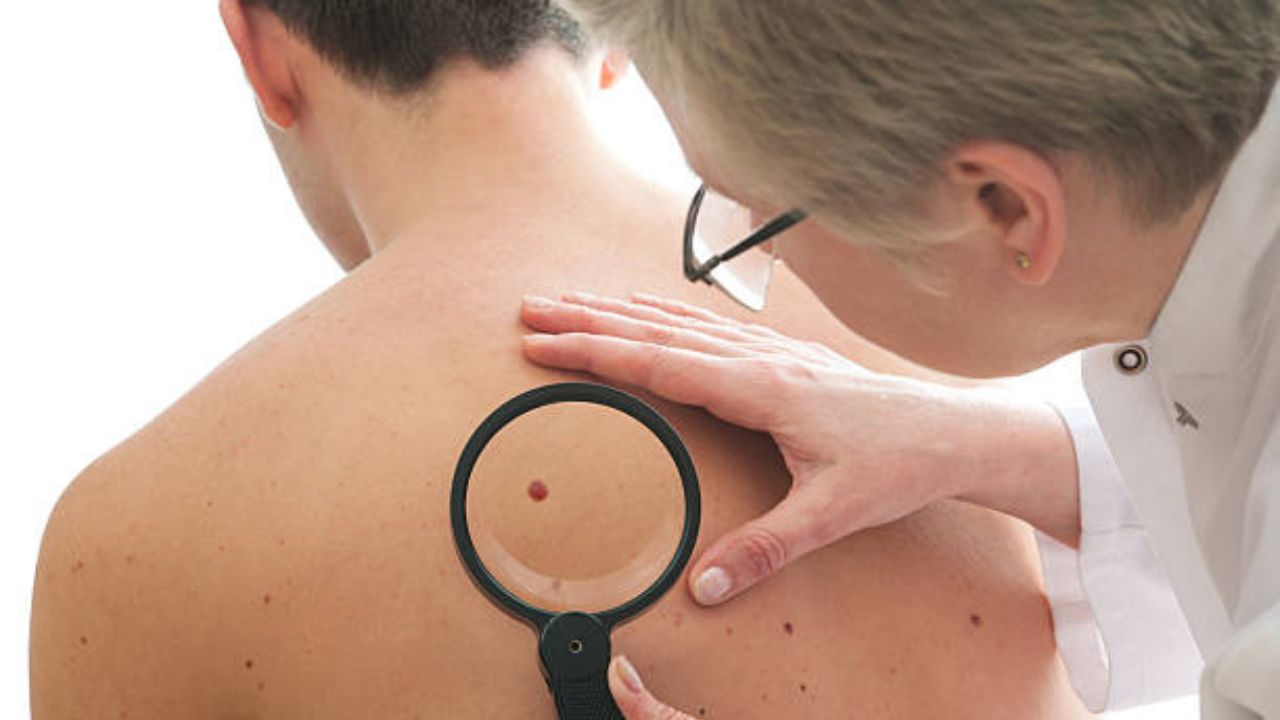
In the next step, the dermatologist examines the conspicuous skin area thoroughly with a reflected light microscope (dermatoscope). He may also want to look at the skin all over his body to see if there are any other noticeable changes.
If the dermatologist suspects skin cancer, he will arrange for further examinations. Above all, this includes taking a tissue sample : the suspicious area of skin is removed under local anesthesia, including a safety margin. The tissue is examined in detail in the laboratory by a pathologist (histologically). In this way, it can be determined whether skin cancer is actually present. In this case, further investigations follow:
- Imaging methods such as X -rays, computer tomography (CT), magnetic resonance imaging (magnetic resonance imaging, MRT) or ultrasound (sonography) can show whether and how far the cancer has already spread in the body (metastasis formation). This is very important for choosing the right therapy.
- Blood tests say something about the general condition of the patient and the function of important organs in the body. This is important, among other things, in order to be able to assess the risk of anesthesia (during the operation).
- Skeletal scintigraphy (bone scintigraphy) is a nuclear medical examination that can be used to detect bone metastases.
Skin cancer screening (early detection)
Statutory health insurance companies in Germany pay for all insured persons over the age of 35 to have a skin cancer screening test every two years. Anyone who has an increased risk can go to free melanoma screening every year. The aim of this completely painless measure is to detect malignant changes in the skin at an early stage. Then the chances of recovery are almost 100 percent.
Skin cancer screening is carried out by a specially trained doctor – i.e. a doctor who has previously undergone a special training program. Skin cancer screening is usually carried out by general practitioners with appropriate training or by dermatologists.
How does skin cancer screening work?
The doctor will first ask you about your general state of health and any previous illnesses and risk factors. Then examine your skin all over your body (including your scalp). He also inspects the external auditory canal as well as the oral mucosa, lips and gums – skin cancer can also develop here.
Before you go for skin cancer screening, you should remove any nail polish from your fingernails and toenails. Skin cancer can also develop under the nails. With color on the nails, however, the doctor could overlook changes.
If the general practitioner is not entirely sure about a skin area, he will refer you to a dermatologist as a precaution. He repeats the skin cancer screening. But this is no cause for concern, because the dermatologist usually gives the all-clear.
The dermatologist takes a closer look at conspicuous skin areas with a special magnifying glass (dermatoscope). If skin cancer is suspected, he may take a tissue sample (biopsy) or completely remove the conspicuous area of skin under local anesthesia and send it to a laboratory. The careful histological examination of the tissue provides certainty as to whether it is actually skin cancer. Very often, however, it turns out that it is just a harmless skin change.
Own contribution to skin cancer prevention
Skin cancer and its precursors can often be detected without expensive tools. Every layman can contribute. It is crucial for your own skin cancer early detection to examine the skin regularly for changes. Do this in a bright, well-lit room, or you may miss minor changes.
Pay particular attention to those areas of the skin that are usually exposed to sunlight, as this is where skin cancer is much more likely to develop. A mirror or an examination by a friend or partner can be helpful for parts of the body such as the back.
It is particularly important in early skin cancer detection to pay attention to changes that could indicate black skin cancer. Keep in mind that this often occurs on parts of the body that are rarely exposed to sunlight.
How can you recognize a malignant melanoma?
The so-called ABCDE rule can be helpful in the early detection of melanoma. It serves as a guide when assessing dark skin spots (birthmarks, pigmented moles):
A (Asymmetry): If a dark patch of skin is not round or oval but is asymmetrical, it should be evaluated by a doctor.
B (boundary): A mole is usually sharply defined from the surrounding skin. If, on the other hand, the spot extends into the surrounding skin or appears blurred, washed-out, skin cancer could possibly be behind it.
C (Colour): If a patch of skin has different shades of color (e.g. light brown and jet black), this is conspicuous. A skin cancer screening brings certainty.
D (Diameter): Any mole larger than two millimeters in diameter must be observed.
E (Elevation): Elevation is how high a mole or other lesion protrudes above the level of the surrounding skin. If the height is more than a millimeter, it can be an indication of skin cancer.
When should you go to the doctor?
Self-examination is very important for early skin cancer detection. If in doubt, you should see a doctor as soon as possible. A doctor’s visit is strongly recommended in the following situations:
- A pigment spot is conspicuous according to the criteria of the ABCDE rule or changes in shape, color or size.
- A pigment spot starts to burn, itch or bleed.
- Rough patches of skin or crusts form. This can indicate white skin cancer. Particular attention should be paid to areas exposed to the sun (face, backs of hands, etc.).
- A new skin lesion (spot, scab) develops in adulthood that does not heal within a few weeks.
- If you have an above-average number and/or irregular pigment spots (birthmarks) (if you have more than 40 or 50 pigment spots, you should see your doctor regularly even if there are no suspicious changes).
- You notice whitish areas / thickening on the lower lip or in the mouth – (pipe) smokers in particular should pay attention to this warning sign.
Skin cancer: disease course and prognosis
The course of the disease depends, among other things, on what type of skin cancer it is. While some tumors only grow slowly, others spread quickly and form metastases at an early stage. This makes it difficult to treat skin cancer. The chances of recovery also deteriorate as the tumor grows and spreads.
In general, the chances of a cure for skin cancer are better the earlier the malignant tumor is discovered and treated. Below you will find more detailed information on the course of the most important forms of skin cancer, chances of recovery and prognosis.
Basal cell cancer
Basal cell cancer grows slowly. In addition, it usually does not cause colonization (metastases) in other organs. This also applies to very advanced tumor stages, when large areas of skin (e.g. the entire nose) have been destroyed by the cancer. Overall, basal cell carcinoma therefore has a good prognosis: it is easy to treat , so that up to 95 percent of patients recover completely. Deaths are rare: only about one in 1,000 patients dies from the cancer.
Risk of recurrence: Regular follow-up visits after completing treatment are very important. In more than four out of ten patients, further basal cell carcinomas form within the first three years after the initial diagnosis. If the check-ups are carried out conscientiously, these new tumors can be detected and treated at an early stage. The intervals at which follow-up examinations make sense depend on the individual case – but one check-up per year is often sufficient. The attending doctor will suggest suitable appointments for each patient. Experts are currently recommending that this aftercare should not be limited in time.
Squamous cell cancer
Squamous cell cancer grows more aggressively than basal cell cancer. It gradually destroys the surrounding tissue. If left untreated, spinalioma can spread throughout the body and form metastases in lymph nodes and other organs. This influences the prognosis: If squamous cell cancer is discovered before it has formed metastases, it is usually easily treatable. As soon as daughter settlements occur, the chances of recovery decrease. Statistically, the cancer is fatal in 40 to 50 out of 1,000 patients.
Risk of recurrence: About half of the patients develop a new tumor within five years after the initial diagnosis. Therefore, regular follow-up examinations should be particularly important in these years. The intervals at which the checks make sense depend on the individual case. In the first year, however, it is advisable to have follow-up visits every three months.
Black Skin cancer
The different types of melanoma show different courses: Some types of melanoma grow on the surface of the skin for a long time and can therefore be treated quite well. Other types penetrate quickly into deeper layers of tissue and soon spread through the blood and lymphatic system in the body. Metastases form early on. The affected patients can die within a few months if left untreated.
Apart from the type of melanoma, the tumor stage at the time of diagnosis also influences the chances of recovery. In Europe, malignant melanomas are often detected at an early stage. Most of the affected patients can be cured. However, the later black skin cancer is discovered and treated, the worse the chances of recovery and the higher the risk of death.
Risk of recurrence: Anyone who has ever had black skin cancer has an increased risk of developing melanoma again (second tumor). That is why regular check-ups after the treatment is over are very important. This follow-up care for melanoma should extend over at least ten years. In the first five years, the check-ups are usually quarterly or semi-annually, after that at longer intervals. However, the doctor treating you will create an individually tailored aftercare schedule for each patient.