Gallstones are crystallized components of the bile. They can develop in the gallbladder or in the bile duct, favored by factors such as obesity and the female gender. Gallstones usually do not cause any symptoms. Depending on the location and size, they can also cause pain – from moderate discomfort in the right upper abdomen to severe biliary colic. Read more about signs of gallstones, treatments for gallstones, diet tips, and prognosis here!
ICD codes for gallstones: K80
Brief overview
- What are gallstones? Crystallized components of the bile in the form of tiny stones (grit) or larger stones. Depending on the location, a distinction is made between gallbladder stones and bile duct stones. Women are more likely to have gallstones than men.
- Risk factors : mainly female, overweight (fat), fertile (fertile), 40 years and more (forty), light-haired (fair), familial disposition (family).
- Symptoms : No or more or less severe symptoms, depending on the location and size of the gallstones, e.g. pain in the right upper abdomen up to severe biliary colic, inflammation of the gallbladder (cholecystitis), biliary congestion with inflammation of the bile duct (cholangitis), jaundice and / or inflammation of other organs.
- Possible consequences : inflammation of the pancreas (acute pancreatitis); Injury to the gallbladder wall with leakage of bile into the abdomen and the resulting peritonitis ; increased risk of gallbladder and bile duct cancer.
- Treatments for gallstones : surgery, medication, shock wave therapy.
Gallstones: description
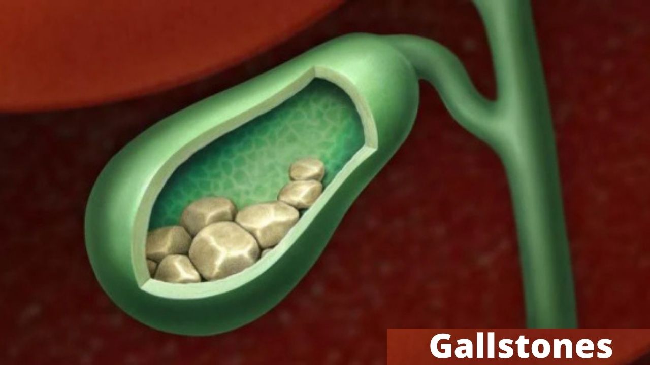
Gallstones also known as cholelithiasis are crystallized components of bile (short: Gall). This fluid is produced in the liver and collected in the gallbladder just below, which is only a few centimeters long. If necessary, the bile is passed through the bile duct into the small intestine, where it helps digest fat.
The main component of bile is about 80 percent water. In addition, there are bile acids, proteins and bilirubin (yellowish breakdown product of the red blood pigment hemoglobin).
The bile also contains cholesterol. Both bilirubin and cholesterol can crystallize – the result is the finest stones a few millimeters in size or gallstones up to several centimeters in size. Doctors then speak of cholelithiasis.
Types of gallstones

Depending on which substance predominates in the gallstones, doctors differentiate between the following two main groups:
- Cholesterol stones : These consist mainly of cholesterol and are responsible for around 80 percent of all gallstone diseases in Germany.
- Bilirubin (pigment) stones: They consist of a cholesterol core to which bilirubin has attached. Bilirubin stones cause about 20 percent of gallstone disease.
Another distinguishing criterion is the location of the cholelithiasis. A distinction is made between:
- Gallbladder stones (cholecystolithiasis): They arise in the gallbladder, the reservoir for the bile.
- Bile duct stones (choledocholithiasis): They are located in the duct connecting the gallbladder and the small intestine. Sometimes they are created on site. Often, however, it is actually gallbladder stones that have been washed out into the bile duct (secondary bile duct stones).
Frequency of gallstones
An estimated 5 to 25 percent of the population have gallstones. Women are two to three times more likely to be affected than men. In addition, the risk of gallstones increases significantly from the age of 40.
Many sufferers do not even know that they have gallstones because they do not (yet) cause symptoms.
Causes and risk factors of gallstones
Gallstones occur when the bile changes in such a way that less soluble components such as cholesterol or bilirubin flocculate. Then tiny crystals form, which combine over time and continue to grow – to form semolina or gallstones.
For most of those affected, several factors contribute to the formation of gallstones (multifactorial genesis). Only very rarely is there a single trigger (such as a genetic defect that inevitably leads to gallstone formation).
Risk factors of the 6-f rule
Certain risk factors promote the development of gallstones. The most important factors can be summarized in the so-called 6-F rule:
- f – female (female)
- f – fat (overweight)
- f – fertile (fertile, several children)
- f – forty (Age 40 years or more)
- f – fair (blonde, light-haired)
- f – family (familial disposition)
The fact that gallstones occur more frequently in some families suggests the influence of genetic factors : for example, a certain variant of the ABCB4 gene can increase the risk of gallstones.
This gene contains the blueprint for a molecular pump that transports cholesterol from the liver cells into the biliary tract. In carriers of the mentioned gene variant, the composition of the bile is changed in such a way that gallstones form more easily.
Very rarely there is a genetic defect that in any case leads to the formation of gallstones.
Other risk factors
Other risk factors for developing gallstones are:
- Pregnancies
- Taking female sex hormones, for example as a contraceptive (pill) or as hormone replacement therapy during menopause
- Certain other drugs such as ceftriaxone (an antibiotic) or somatostatin (for the hormone disorder acromegaly or for bleeding in the upper digestive tract)
- Bile congestion with impaired mobility of the gallbladder (if the gallbladder cannot contract properly, the bile builds up and gallstones form more easily)
- Bile acid loss syndrome (disease with a relevant deficiency in bile acids, e.g. as a result of the surgical removal of a large part of the small intestine – for example in Crohn’s disease)
- Diabetes mellitus
- Cirrhosis of the liver (e.g. due to high alcohol consumption)
- Increased levels of fat in the blood (triglycerides, cholesterol)
- Severe overweight (obesity)
- Severe weight loss within a short time, e.g. with a weight reduction diet (diet in which less energy is absorbed than the body actually needs) or surgical stomach reduction (for people who are very overweight)
- Special, high-calorie tube feeding
The fact that women get gallstones more often than men is probably due to the female sex hormones. This is also supported by the fact that taking such hormones (e.g. as a contraceptive pill) and pregnancy increase the risk of cholelithiasis.
Symptoms of gallstones

Gallbladder stones symptoms : most people with gallstones do not experience any discomfort. This is called “silent” gallstones. If at all, they are usually only discovered by chance, for example as a secondary finding in an ultrasound or X-ray examination.
Sometimes “silent” stones become “speaking” over time and thus begin to cause discomfort. Studies have shown that two to four in 100 people with gallstones develop noticeable symptoms within a year.
Symptomatic gallstones are gallstones that cause discomfort. These can be very different. In milder cases, pain and unspecific complaints occur in the upper abdomen, such as feelings of fullness or pressure, belching and flatulence. These symptoms usually show up after a meal and can be exacerbated by the consumption of fatty and / or fried foods.
Sometimes gallstones also trigger biliary colic – severe, cramp-like pain in the right middle and upper abdomen. They are wave-like : the pain swells quickly, then reaches a plateau and then subsides spontaneously or after taking medication.
In addition, biliary colic typically lasts 15 minutes to several hours. Sometimes the pain radiates to the back and right shoulder region. In addition, accompanying symptoms can include sweating, nausea, nausea and vomiting.
Biliary colic (biliary pain) occurs mainly at night and often not in chronological order after a meal.
Around every second patient who has had gallstone symptoms such as colic at some point will experience symptoms again within two years.
The size and location of the gallstones are decisive
Whether or not gallstones trigger symptoms depends, among other things, on their size. Most are rather small, like a cherry or a hazelnut, and often do not cause any discomfort. Others reach the size of a hen’s egg. Then pain is very likely.
The location of the cholelithiasis also influences the extent to which symptoms occur. Basically, symptoms are observed more often with bile duct stones than with gallbladder stones. They trigger colic-like pain when they get stuck in the bile duct and block it – the bile can then no longer drain into the small intestine.
Gallbladder stones cause colic when the stones block the exit of the gallbladder into the bile duct or its opening into the duodenum. Again, the gallbladder fails to force bile into the small intestine, which leads to intensified, painful contractions.
Doctors refer to the build-up of bile as a result of an obstruction to drainage as bile build-up (cholestasis).
Complications of gallstones
Gallstones can have several effects:
Inflammation of the gallbladder and possible consequences
When gallbladder stones block the gallbladder exit, the bile builds up in it. This can lead to acute inflammation of the gallbladder (cholecystitis): the accumulated bile overstretched the gallbladder wall, and the mucous membrane lining the organ becomes irritated and inflamed.
Bacteria can then settle on it more easily. Signs of inflammation of the gallbladder include severe pain in the upper abdomen, fever, and chills.
If left untreated, acute gallbladder inflammation can lead to the formation of pus in the gallbladder (gallbladder empyema) – possibly even with partial death and thus rupture of the gallbladder wall (gallbladder perforation). In extreme cases, the peritoneum can become inflamed (“bilious” peritonitis = “bilious” peritonitis).
The inflammation can also spread to the whole body via the blood – doctors then speak of ” blood poisoning ” (sepsis).
Sometimes the inflammation of the gallbladder is chronic rather than acute. In very rare cases, the gallbladder wall can thicken and calcify as a result – doctors speak of this as ” porcelain gallbladder “. The organ can then no longer contract properly. A certain form of the “porcelain gallbladder ” also increases the risk of gallbladder cancer.
Inflammation of the biliary tract and jaundice
When a gallstone blocks the bile duct, the bile builds up in it. The possible consequence is an inflammation of the bile ducts (cholangitis) with severe upper abdominal pain, fever and chills.
In addition, the inflammation can lead to jaundice (jaundice): Because the bile congestion means that the breakdown product of the red blood pigment – yellow bilirubin – can no longer be excreted, it is deposited in the tissue.
Especially the “white” in the eyes and the skin can turn yellow. In addition, the stool turns light and the urine dark.
Like gallbladder inflammation, bile duct inflammation can also spread to neighboring organs.
Inflammation of the pancreas
In most people, the bile duct, along with the duct of the pancreas (pancreas), joins the duodenum – the uppermost section of the small intestine. If a gallstone blocks the opening in the intestine, the secretion of the pancreas can also build up.
The possible consequence is inflammation of the pancreas (acute pancreatitis) with severe epigastric pain, nausea, vomiting and fever.
The acute pancreatitis often resolves spontaneously. However, the same applies here: the inflammation can spread to neighboring organs.
Gallbladder and bile duct cancer
Gallstones increase the risk of gallbladder cancer and bile duct cancer – but only slightly. In addition, both types of cancer are rare.
However, people with a certain form of the very rare porcelain gallbladder mentioned above are very susceptible to gallbladder cancer. It is therefore usually recommended that you have your gallbladder removed as a precaution.
Examinations and diagnosis of gallstones
If you are suspected of having gallstones, the doctor will first take a detailed discussion about your medical history (anamnesis). Among other things, he asks you to describe all complaints in detail. He also asks about any previous or underlying illnesses. This is followed by a comprehensive physical exam and imaging tests.
Imaging procedures
The most important imaging for suspected gallstones is the ultrasound examination (sonography) the abdomen. In most cases, gallbladder stones from a size of one to two millimeters can be detected.
In addition, the doctor can recognize any other pathological changes in the ultrasound image. In the case of a gallbladder inflammation, for example, the gallbladder wall is thickened and layered.
However, bile duct stones cannot always be detected with the help of a conventional ultrasound examination (via the abdominal wall). Endosonography – an ultrasound examination from the inside – achieves a better hit rate here.
The doctor guides a thin, flexible tube with an ultrasound head through the mouth, esophagus and stomach and into the duodenum to the confluence of the biliary and pancreatic ducts. Any bile duct stones can be seen well through the wall of the duodenum.
A special X-ray examination, the endoscopic retrograde cholangio-pancreatography (ERCP), can also clearly identify gallstones in the gallbladder and bile duct. In addition, smaller stones can be removed immediately during the examination.
Another imaging technique that can be used to clarify gallstones is magnetic resonance cholangio-pancreatography (MRCP). This is an examination of the biliary tract and the pancreatic duct using magnetic resonance imaging (magnetic resonance imaging, MRI).
Blood test
In addition to the imaging tests, blood tests are important. Abnormal values can indicate complications caused by the gallstones. If, for example, gamma-GT and / or alkaline phosphatase (AP) are increased, this can indicate a disease of the biliary tract.
Bilirubin levels are typically increased when a gallstone blocks a larger biliary tract (obstructive jaundice). Increased white blood cell (leukocyte) readings and blood sedimentation (sedimentation rate) may indicate inflammation of the gallbladder or bile ducts.
Further examinations if necessary
Sometimes gallstones occur under unusual conditions – for example in families, already in childhood or adolescence, or repeatedly in the bile duct. Then further examinations should clarify the exact cause. For example, if a certain genetic cause is suspected, a genetic analysis can help.
Treatments for gallstones

Whether treatments for gallstones is necessary depends on where the stones are located and whether and what symptoms (such as biliary colic) they are causing.
Therapy of biliary colic
The doctor treats acute biliary colic with anticonvulsant and pain reliever drugs (antispasmodics and analgesics) such as ibuprofen.
If the gallbladder becomes infected, the patient is also given antibiotics. In the first 24 hours after the onset of biliary colic, the patient must also not eat any food (zero diet).
If you have an acute biliary colic that lasts for several hours and is associated with very severe symptoms, you should call the emergency doctor!
Therapy of gallstones
Gallbladder stones usually only need treatment if they cause symptoms or complications such as gallbladder inflammation. Bile duct stones, on the other hand, should always be treated as they often lead to complications.
Remove gallstones
There are several methods of removing gallstones. Which procedure is used depends, among other things, on the location (gallbladder or bile duct) and the size of the gallstones.
Gallstones are usually removed surgically . This is usually done as part of a so-called laparoscopy. However, other surgical techniques are also available.
In the case of repeated attacks of pain and acute inflammation of the gallbladder, the gallbladder is generally removed during the procedure (cholecystectomy). The body then stores the bile in the bile duct.
In certain cases, an alternative to surgery is medical treatment of the gallstones. The patient has to take a preparation that can dissolve the stones over a longer period of time. In addition, gallstones can also be smashed with the help of shock waves (shock wave therapy).
Diet for gallstones
With the right diet, you can prevent gallstone problems and also prevent the formation of (new) gallstones. To do this, you should eat as little fat as possible: Dietary fat promotes biliary colic and stone formation.
In addition, you should eat a wholesome and high-fiber diet. Include whole grains, vegetables and fruits on your diet regularly. This diet – in combination with regular exercise and sport – can help maintain a healthy body weight or reduce excess fat. Obesity is one of the most important risk factors for gallstones.
Gallstones foods to avoid :
- Refined breads, pastas, etc.
- High-fat dairy.
- Vegetable oil.
- Peanut oil.
- Processed foods.
- Sugar.
- Alcohol.
Course and prognosis of gallstones
Gallstones, which are causing discomfort, are generally quite easy to remove. Surgery has the best prognosis. The gallbladder is often removed as well.
Afterwards, relapses are relatively rare (with gallstone formation in the bile duct). With non-surgical treatment, the relapse rate is higher.