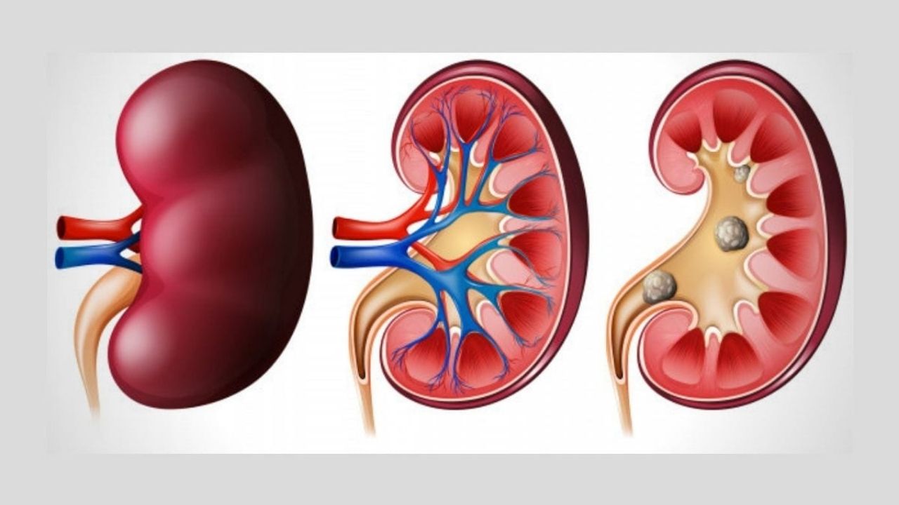
Renal colic is a condition caused by a violation of the outflow of urine from the cavity system of the kidney, manifested by a violation of the organ and severe pain syndrome.

A few words about anatomy and physiology
The kidneys are filters of the human body with a huge number of precise settings. They are approached by arteries that carry blood under high pressure. Once in the kidney tissue, blood is filtered through special semi-permeable membranes, so that all substances unnecessary for the body (for example, protein breakdown products, so-called “nitrogenous waste”, in particular, urea or urea) enter the urine and are excreted from the body. and useful substances – glucose, proteins, minerals – are reabsorbed and returned back into the bloodstream. Naturally, the renal filter does not allow blood cells (erythrocytes, leukocytes, etc.) to pass into the urine. After urine has formed, it enters the collecting duct and tubule system (passing through the renal columns and renal papillae), which open into the renal cavity system.
From the renal pelvis urine through a thin (no more than 7-8 mm) tube – the ureter – enters the bladder. Moreover, at the junction of the ureter with the bladder there is an antireflux mechanism – a kind of system – a nipple. Urine can flow from the ureter to the bladder, but it cannot go in the opposite direction. This helps to protect the kidney from urinary backlash when the pressure in the bladder rises. As the bladder fills, the urge arises and the person urinates through the urethra.
How does renal colic develop?
Normally, the pressure in the renal pelvis does not exceed 7-10 cm of water. This is a very low pressure. And the kidney can work normally only with these indicators. If the pressure in the renal cavity system for some reason begins to grow, a number of serious problems develop.
Renal colic is the result of a rapid blockage (obstruction) of the urine flow. At the same time, urine continues to be produced for some time, filling the entire cavity system of the kidney to failure – “hydronephrosis” or “dropsy of the kidney” develops. After that, the production of urine stops – the kidney stops working normally. In such a situation, conditions are created for the development of many life-threatening problems, for example, acute inflammation of the kidney – acute pyelonephritis, which can quickly lead to sepsis and death of the patient.
The main causes of renal colic are:
- Blockage of the ureteral lumen with a stone (ureteral stone) is the most common cause of renal colic. The stones form in the renal calyx, after which they can migrate downward in the direction of the pelvis and, then, the ureter. The latter, as already mentioned, usually has a diameter of no more than 8 mm. In addition, there are physiological contractions. It is important to note that gender, height and weight of a person do not allow predicting the diameter of the ureter. It is not uncommon for large adult males to have a thinner ureter than in miniature females. When a stone gets stuck in the ureter, swelling and inflammation develops in the wall of the latter, which blocks the stone, preventing it from moving on.
- Blockage of the exit from the pelvis into the ureter (kidney stone) – a similar problem usually occurs with relatively medium- sized (up to 1.5-2 cm) kidney stones, which, like a valve, are able to close the anastomosis of the pelvis and ureter. This variant of renal colic can self-relieve when the position of the body changes (when the stone “falls off” from the exit into the ureter).
- Bleeding from the upper urinary tract. In this situation, the ureter becomes clogged with blood clots. As a rule, the cause of this condition is bleeding from a tumor of the cavity system of the kidney and ureter.
- Iatrogenic (associated with medical manipulation) damage. This type of renal colic usually develops after surgery on the pelvic organs.
Renal colic signs
- severe pain syndrome in the lumbar region with spread to the lateral parts of the abdomen, groin (depending on the localization of the obstacle to the outflow of urine); at the same time, there is no such position in which the pain subsides, the patient “does not find a place for himself”,
- nausea and vomiting that does not bring relief,
- bloating
- pallor of the skin,
- dry mouth.
Help with renal colic
At the prehospital stage, renal colic can be relieved with pain relievers and antispasmodics. In particular, diclofenac (voltaren) can be considered the best option. It can be injected into a muscle or administered as a rectal suppository – 100 mg. But no more than 300 mg per day. Pill formulations are usually poor at relieving pain in renal colic.
Severe renal colic is superior to labor on a pain scale. Therefore, this condition sharply reduces the person’s capacity to function.
Pain relief is a temporary measure. It is necessary to contact a specialized hospital as soon as possible.
If the hospital in which the patient got to works according to modern standards, which are clearly formulated in the European recommendations for urology and has the necessary equipment, then the effective treatment of uncomplicated renal colic in most cases is a matter of one-day hospitalization without “adventures”. And on the contrary, in dense conditions, in the absence of the necessary knowledge, equipment, supplies, etc., a banal situation can turn into an extremely serious problem, up to the loss of a kidney or life-threatening complications.
At the first stage, the patient is diagnosed. The modern approach in this case is a non-contrast low-dose computed tomography (CT) of the kidneys and urinary tract. Ultrasound and intravenous urography (conventional x-rays) do not provide enough information to plan for safe and effective treatment.
With a stone size of more than 5-7 mm, the likelihood of its independent withdrawal without the emergence of new problems is unlikely. In such cases, it is advisable to resort to active treatment tactics. As a rule, at the first stage, a drainage intervention is performed, that is, the outflow of urine from the kidney is improved. This can be done in two ways:
1. Installation of an internal ureteral stent.

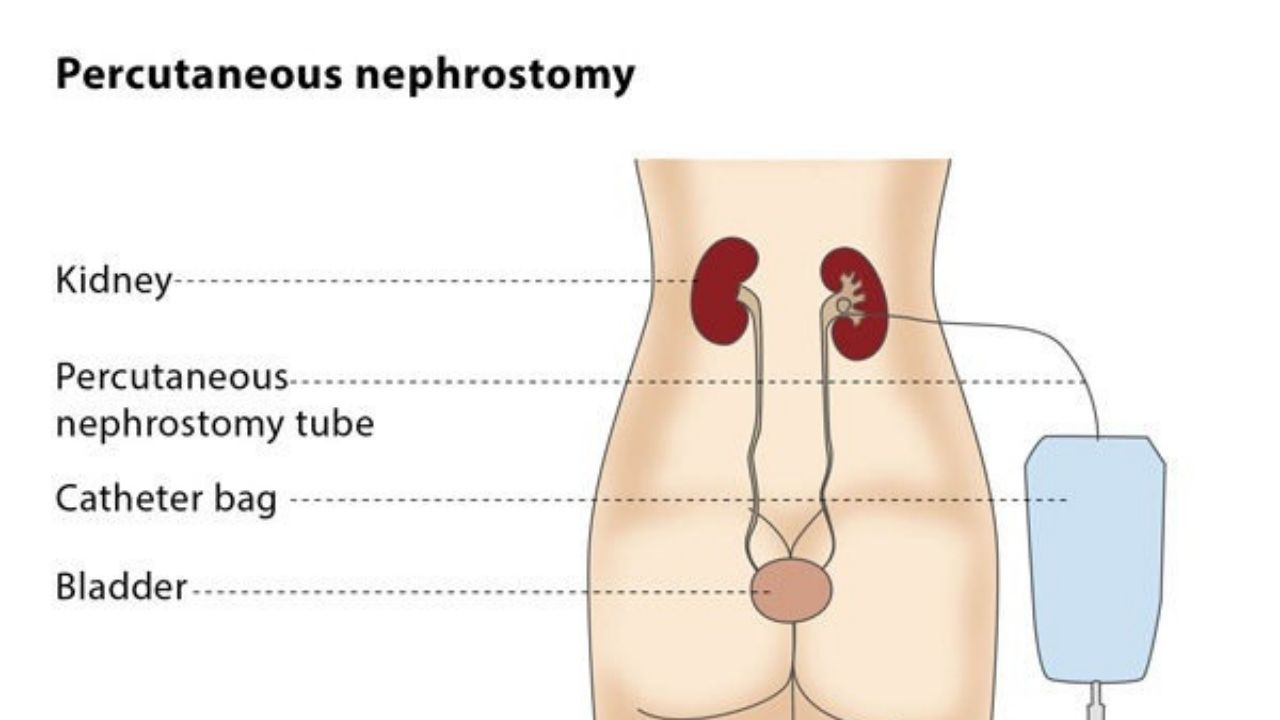
2. Performing percutaneous nephrostomy, which is extremely rare.

If the kidney is adequately drained, it starts to work, the risk of infection, sepsis, etc. becomes minimal and all that remains is to remove the stone at the second stage of treatment (and, of course, remove the drainage devices). In some cases, it is advisable to immediately remove the stone without draining. However, such operations are often more traumatic and can cause additional problems (for example, trauma to the ureter). Therefore, in most cases, it is advisable to avoid them. Most patients with an internal ureteral stent inserted can return to their normal activities and even return to work.
After 7-14 days, the second stage of treatment is usually carried out – in fact, crushing of the ureteral stone. This is done with a special thin endoscope using laser, ultrasound, pneumatic energy, or simply using special forceps or extractor baskets. Laser crushing of ureteral stones has certain advantages over other types of energy. The main one is that a laser is capable of destroying a stone of almost any density. But in the right hands, ultrasound and pneumatic energy can provide absolutely equivalent results.
Laser crushing is possible with flexible endoscopes (for kidney stones), since the thin laser fiber bends over a wide range. But there are other options for lithotripsy using flexible probes, for example, electrical impulse. Standard stone removal after previous stenting is also performed in a hospital for one day (or one day).
In cases of large (or coral- shaped) kidney stones leading to renal colic, other treatments are used, such as percutaneous (percutaneous) lithotripsy using ultrasound, pneumatics, or laser.
In conclusion, it should be noted that effective and safe treatment of urolithiasis is possible only in hospitals where all three essential conditions are met: personnel trained in accordance with modern standards, the necessary equipment and high-quality consumables.